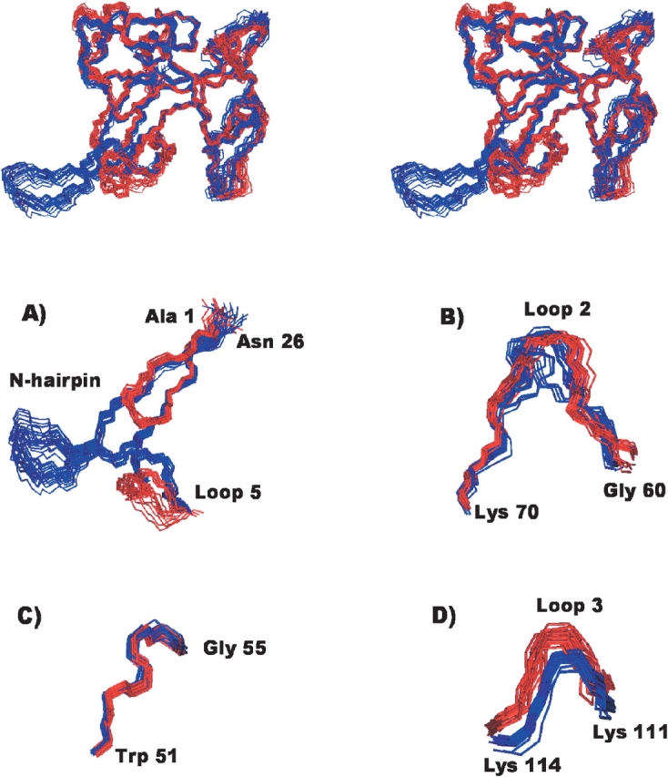
Figure 2.

Structural comparison of the wild-type α-sarcin and the Δ(7-22) α-sarcin deletion mutant. (Top) Stereoscopic view of the superposition of the two ensembles of their 3D NMR structures; the 20 conformers of the wild type α-sarcin are in blue, and those of the Δ(7-22) mutant, in red. (Bottom) Detail of the superposition of the backbone atoms corresponding to some regions of the wild-type α-sarcin (blue) and the Δ(7-22) α-sarcin deletion mutant (red) structures. (A) Relative orientation of the N-terminal β-hairpin and loop 5, (B) segment of loop 2, (C) residues 51–55, and (D) lysine-rich fragment of loop 3.
