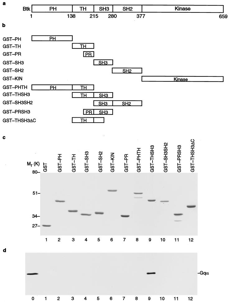
Figure 1.
Gqα association with Btk. (a) Schematic representation of Btk and its structural domains. Kin, kinase domain. The numbers at the bottom indicate positions of amino acid residues. (b) Representation of GST-fusion protein constructs. (c) Coomassie staining of SDS/PAGE of purified GST-fusion proteins. (d) Binding of Gqα to the THSH3 region of Btk. GST-fusion proteins were incubated with whole-cell extracts made from Gqα(Q209L)-transfected HEK293 cells. After SDS/PAGE, bound Gqα was detected with anti-Gqα antibody. Lane 0 was loaded with 40 μl of whole-cell extract as control. Lanes 1–12 are loaded with GST-fusion proteins as indicated in c.

