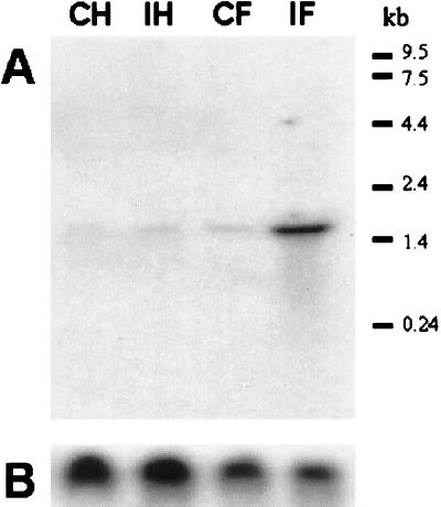
Figure 7.
Northern blot analysis of PAP mRNA level. RNA samples (10 μg) from hemocytes of control insects (CH), hemocytes collected 24 h after injection of E. coli (IH), fat body from control insects (CF), or fat body collected 24 h after injection of E. coli (IF) were separated by electrophoresis on a 1% agarose gel containing formaldehyde. The RNA was transferred to a nitrocellulose membrane and then hybridized with 32P-labeled M. sexta PAP cDNA (A). The positions of RNA standards are shown at the side of each blot. Approximately equal RNA loading of each lane was confirmed by probing a duplicate blot with 32P-labeled cDNA for M. sexta ribosomal protein S3 (B) (29).

