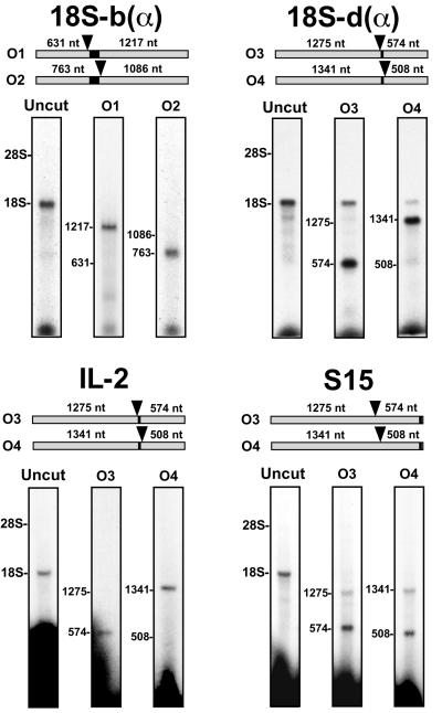
Figure 4.
Mapping the binding sites of four RNA probes by RNase H digestion of the rRNA. 18S-b(α) was localized by using oligonucleotides O1 and O2 to direct RNase H digestion. O1 is complementary to nucleotides 632–652 and O2 is complementary to nucleotides 764–783. 18S-d(α) was localized by using oligonucleotides O3 and O4. Oligonucleotide O3 is complementary to nucleotides 1276–1295, and O4 is complementary to nucleotides 1342–1361. Il-2 and S15 were localized by using oligonucleotides O3 and O4. In the upper section of each panel, the two gray bars represent the 18S rRNA. The black bar indicates the position of complementarity to the probe, and the oligonucleotides (O1-O4) used for the RNase H digestions are represented as arrowheads. The sizes of the rRNA fragments expected with each oligonucleotide are indicated above the 18S rRNA bar. The lower section of each panel shows the results of the RNase H digestions. After cross-linking the probes to ribosomes, as described in Materials and Methods, the RNAs were purified away from protein, annealed to one of the oligonucleotides, and the rRNA at this position was digested with RNase H. The positions of the rRNAs were determined by ethidium bromide staining of the agarose gels.

