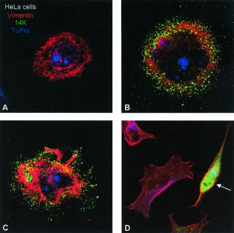
FIG. 2.
Differential morphological changes induced in epithelial HeLa cells after infection with the WR and MVA VV strains. HeLa cells were infected with WR and MVA at 0.05 PFU/cell, and at 48 hpi cells were fixed with paraformaldehyde, glutaraldehyde, and Triton X-100 in CB. Soluble cytoplasmic p14 viral protein and virions were detected with an antibody to the p14 protein (A27L gene) of VV and a secondary antibody conjuged with FITC; the cellular cytoskeletal vimentin was detected with a monoclonal antibody and a secondary antibody conjugated with TRITC (tetramethyl rhodamine isothiocyanate); the DNA was visualized by staining with To-Pro. More than 50 fields at ×40 magnification were captured, and the changes in the morphology of infected cells were registered and quantified (not shown). (A) Mock-infected cell. (B and C) Cells infected with WR, exhibiting a rounded and multipolar morphology. (D) MVA-infected cell with a characteristic bipolar morphology (arrow).

