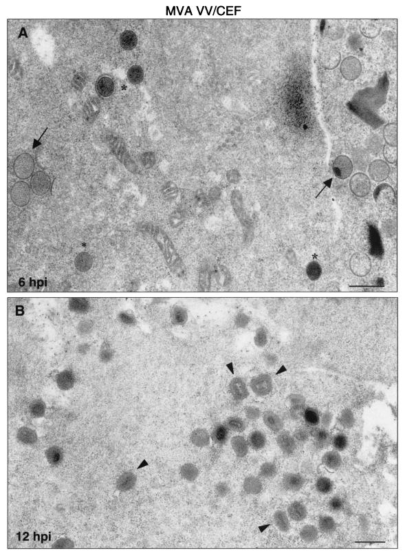
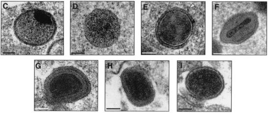
FIG. 7.
Morphogenetic events in CEFs infected with MVA. Primary CEFs infected at an MOI of 5 PFU/cell were chemically fixed at 6 and 12 hpi and processed for conventional embedding in an epoxy resin. Ultrathin sections were studied by transmission electron microscopy at low and high magnifications. (A) At 6 hpi, IVs (arrows) and spherical and dense transition forms (asterisks) were abundant. (B) At 12 hpi, IMVs constituted the most abundant viral form (arrowheads). Also, a DNA crystalloid is seen at 6 hpi. The hypothetical sequence of morphogenesis of the MVA is shown in panels C to I at higher magnifications. (C) Characteristic IV with DNA spot; (D) transition IV→IMV virus; (E) viral form occasionally seen wrapped by two membrane units (arrowhead). In this sequence the traditional stages of maturation of VV are also shown: IMV (F), IEV (G), and EEV (H). (I) Finally, the “transition viral form” IV→IMV is frequently seen out of the cell. Bars: 0.5 μm, A and B; 100 nm, C to I.


