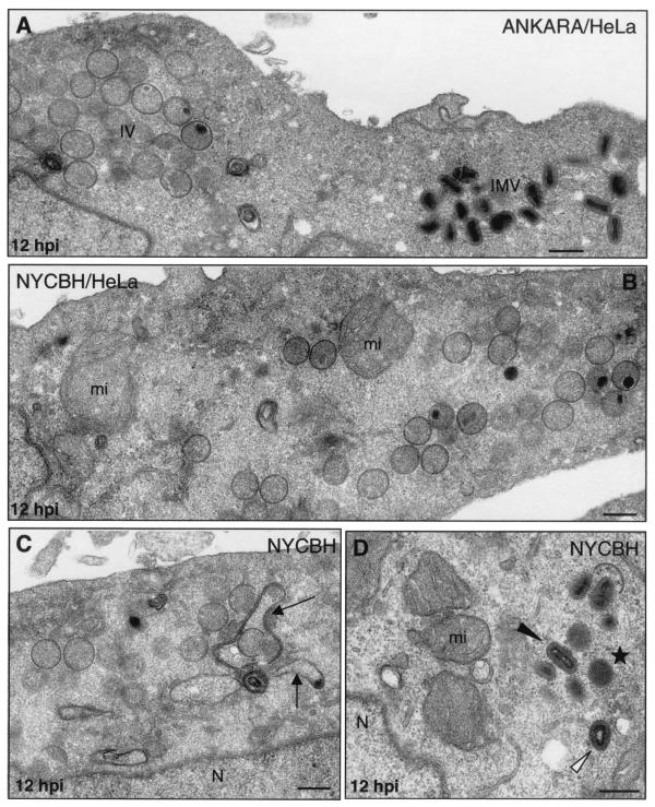
FIG. 9.
Assembly of Ankara and NYCBH strains of VV in HeLa cells. HeLa cells were infected with 5 PFU of Ankara or NYCBH strains of VV/cell, and at 12 hpi the cells were processed for electron microscopy analysis. (A) Both IVs and IMVs accumulated in the cytoplasm of HeLa cells infected with Ankara strain. (B) The NYCBH strain accumulated large amounts of IVs in the perinuclear areas of HeLa cells, where numerous swollen mitochondria (mi) are seen. (C and D) In these cells IVs are frequently surrounded by dense membranes (marked by arrows in panel C), and small groups of IMVs (black arrowhead in panel D) close to dense “transition” viral particles (star, panel D) and “atypical IMVs” (white arrowhead in panel D) are frequently seen. N, nucleus. Bars, 300 nm.

