Figure 10.
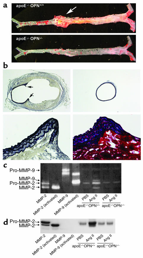
Decreased Ang II–induced abdominal aortic aneurysm formation and MMP-2 and MMP-9 activity in ApoE–/–OPN–/– mice. (a) Representative aorta from ApoE–/–OPN+/+ and ApoE–/–OPN–/– mice infused with Ang II for 4 weeks. The arrow indicates a typical abdominal aortic aneurysm in ApoE–/–OPN+/+ mice. (b) Characteristics of aneurysmal tissue from the abdominal aorta of ApoE–/–OPN+/+ and ApoE–/–OPN–/– mice infused with Ang II. EVG staining of the elastic lamina (black) of ApoE–/–OPN+/+ (upper left) and ApoE–/–OPN–/– (upper right) mice (both ×20 magnification). The arrows indicate the disruption of the media and breaks of the elastic lamina. EVG (lower left) and trichrome (lower right) staining at higher power demonstrate the abdominal aortic wall of ApoE–/–OPN+/+ mice containing smooth muscle cells (red) between elastic fibers (black) at the edge of the disrupted media. (c) Representative zymography analysis of MMP activities in the aorta from Ang II–infused ApoE–/–OPN+/+ and ApoE–/–OPN–/– mice. Recombinant mouse MMP-2 and MMP-9 was activated by incubation with p-aminophenylmercuric acetate and used for calibration. (d) Representative Western blot analysis of aortic MMP-2 expression in ApoE–/–OPN+/+ and ApoE–/–OPN–/– mice. Recombinant MMP-2 and MMP-9 was used as positive control and for determination of specificity.

