Figure 5.
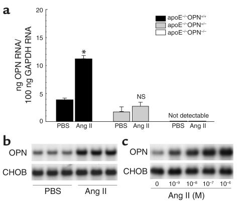
OPN mRNA expression in Ang II–infused mice and murine macrophages. (a) Two weeks after infusion with vehicle or Ang II, OPN mRNA of the thoracic aorta from ApoE–/–OPN+/+ (black bars), ApoE–/–OPN+/– (gray bars) and ApoE–/–/OPN–/– (white bars) mice was analyzed by quantitative real-time RT-PCR. Values are normalized for GAPDH expression and expressed as mean ± SEM. *P < 0.05 versus ApoE–/–OPN+/+ mice infused with vehicle. (b) Two weeks after infusion with vehicle (PBS) or Ang II, peritoneal leukocytes from wild-type control mice (n = 3 in each group) were isolated after intraperitoneal injection of thioglycollate. Total RNA was analyzed for OPN expression by Northern blotting. Blots were cohybridized with cDNA encoding the constitutively expressed housekeeping gene CHO gene B (CHOB) to assess equal loading of samples. (c) Peritoneal macrophages were serum deprived for 24 hours and stimulated with increasing doses of Ang II. Twenty-four hours after stimulation, RNA was isolated and analyzed for OPN expression by Northern blotting. The autoradiograms shown are representative of three independently performed experiments.

