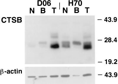
Figure 6.
Western blot analysis of total protein extract from normal squamous esophagus (N), Barrett’s esophagus (B), and esophageal adenocarcinoma (T) specimens probed with a polyclonal anti-CTSB antibody. A 27-kDa doublet band is identified in all specimens. The lower Mr band comprising this doublet is more abundant in tumor specimens. A higher Mr band, associated with extracellular expression of CTSB protein, also is identified in the tumor specimens.

