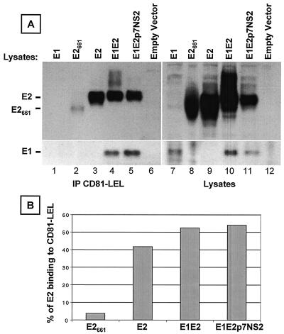
FIG. 1.
Interaction of soluble CD81 with different forms of HCV glycoproteins. (A) Human embryonic kidney 293T cells transfected with a plasmid encoding E1 (lane 1), truncated E2661 (lane 2), E2 (lane 3), E1E2 (lane 4), or E1E2p7NS2 (lane 5) or with an empty vector (lane 6) were lysed at 72 h posttransfection. Cleared lysates (5 × 10⁶ cell equivalents) were precipitated with a recombinant fusion protein containing the LEL of human CD81 fused to glutathione S-transferase (CD81-LEL) preadsorbed onto glutathione-Sepharose 4B beads. Precipitations (lanes 1 to 6) and lysates (lanes 7 to 12; 3 × 10⁵ cell equivalents) were separated by SDS-10% PAGE under reducing conditions, and immunoblots were analyzed with anti-E2 MAb 3/11 (top). The blots were then stripped and reprobed with the anti-E1 MAb, A4 (bottom) as previously described (13). (B) To compare the binding of various forms of E2 to CD81, the intensities of monomeric E2 were measured with ImageQuant 3.3 (Molecular Dynamics). The percentage of E2 binding to CD81 was calculated as follows: [(E2 precipitated by CD81-LEL)/(total E2 in cell lysate)] × 100.

