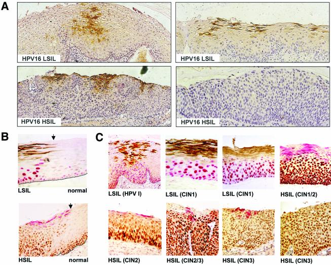
FIG. 8.
Distribution of HPV16 E4 and MCM in precancerous cervical epithelium infected with HPV16. (A) Detection of HPV16 E4 (brown) by immunoenzymatic staining in a region of HPV16 LSIL (upper panels) and HSIL (lower panels). Lesions were counterstained with hematoxylin to enable grading. Images were taken using a 4× objective. (B) Detection of HPV16 E4 and MCM at the edge of an HPV16-induced LSIL (upper panel [brown, E4; red, MCM]) or HSIL (lower panel [red, E4; brown, MCM]). E4 staining is not apparent in normal epithelial tissue. In normal cervical epithelium, MCM expression is confined to cells of the basal and parabasal cell layers. Images were taken using a 10× objective. (C) Patterns of HPV16 E4 and MCM expression in cervical lesions showing some evidence of HPV16 late gene expression. The lesions examined included HPVI and CIN1 (brown, E4; red, MCM) and CIN1/2, CIN2 and CIN3 (red, E4; brown, MCM). The progressive loss of HPV16 E4 staining is accompanied by an increase in the abundance of cells that express MCM. Images were taken using a 10× objective.

