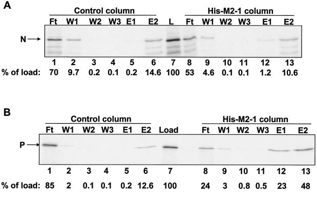
FIG. 1.
M2-1 affinity chromatography with IVT N or P protein. RSV N (A) and P (B) proteins were expressed and labeled with [35S]methionine by IVT. Columns (20 μl each) containing 18.5 μM immobilized M2-1 or control columns containing no immobilized protein were loaded with 20-μl IVT protein lysates. Columns were washed (three times with 40 μl) and eluted first with 40 μl of buffer containing 1 M NaCl (E1) and then with 40 μl of buffer containing 1% SDS (E2). Lanes: L, IVT protein lysate that was loaded onto each column (5 μl per lane); Ft, flowthrough fractions (5 μl per lane); W1 to W3, wash fractions (10 μl per lane). Ten microliters each of E1 and E2 was loaded in each lane. The positions of N and P are indicated to the left of each gel. Phosphorimages of the gels were quantified. The amount of N or P protein present in each fraction was normalized to the amount of the protein in the load sample (percentage of load), as indicated below each gel.

