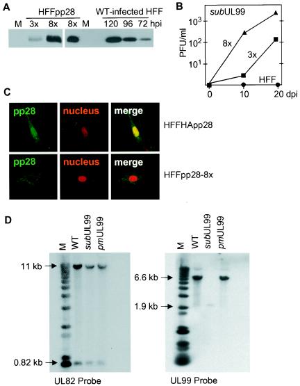
FIG. 2.
Characterization of complementing cells and analysis of the structure of mutant DNAs derived from pp28-deficient virus produced in complementing cells. (A) Western blot analysis monitoring the expression of pp28 in mock-infected (M), HFFpp28-3x cells (3x), HFFpp28-8x cells (8x), and HCMV-infected fibroblasts (HFF) at different times (hours) postinfection (hpi). WT, wild type. (B) Growth of BADsubUL99 in HFFpp28-3x cells (3x), HFFpp28-8x cells (8x), and normal fibroblasts (HFF). Cells were infected, lysates of cells were prepared in the culture medium at the indicated times after infection, and virus was quantified by plaque assay on HFFpp28-8x cells. (C) Location of native pp28 and HA-tagged pp28 expressed from recombinant retroviruses in fibroblasts. Immunofluorescence was performed by using a pp28-specific antibody on HFFpp28-8x (bottom) cells and cells expressing HA-tagged pp28 (HFFHA-pp28, top). Three images are presented for each cell type: left, pp28 (green); middle, nuclear stain (red); right, multicolor merge. (D) Southern blot assay of viral DNAs. Total cell DNA was prepared from HFFpp28-8x cells infected with the indicated viruses, and DNA was cleaved with HindIII and analyzed by Southern blot assay with a UL82- and UL99-specific probe DNA. The sizes of labeled DNA fragments are indicated in kilobases relative to marker (M) DNA fragments.

