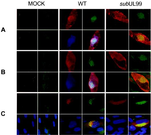
FIG. 8.
Localization of viral proteins expressed in normal fibroblasts infected with wild-type (WT) and pp28-deficient viruses. Cells were infected with BADwt or BADsubUL99 at a multiplicity of infection of 0.01 PFU/cell and processed for immunofluorescence at 120 h postinfection. Four images are presented for each cell. The upper-left quadrant shows UL32-encoded pp150 (A), UL83-encoded pp65 (B), or UL55-encoded gB (C) in red. The lower-left quadrants of panels A and B display pp28 in blue, whereas the lower-left panel of C contains blue-stained nuclei. In all panels, the upper-right quadrant displays Golgi in green and the lower-right panel shows a multicolored merged image.

