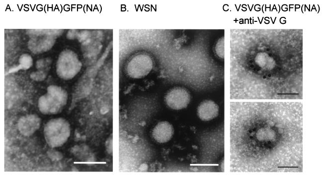
FIG. 5.
Electron microscopy of VSVG(HA)GFP(NA) virus. VSVG(HA)GFP(NA) (A) and WSN (B) viruses were centrifuged through 20% sucrose, and the pelleted materials were then negatively stained with 2% PTA. (C) Pelleted VSVG(HA)GFP(NA) virus was also immunolabeled with anti-VSVG monoclonal antibody conjugated to 15-nm gold particles. Bar, 100 nm.

