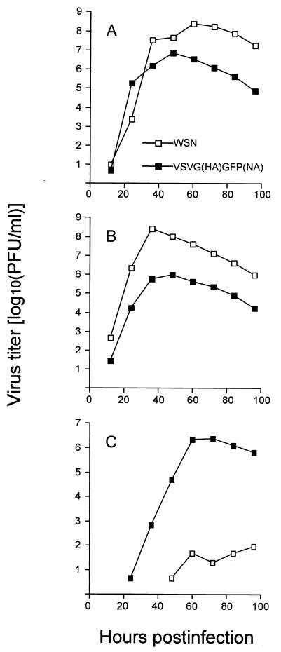
FIG. 6.
Growth curves of VSVG(HA)GFP(NA) virus in BHK, CHO, and MDCK cells. BHK (A), MDCK (B), and CHO (C) cells were infected with virus at a multiplicity of infection of 0.001. At the indicated times after infection, the virus titer in the supernatant was determined by using MDCK cells. The values are means of duplicate experiments.

