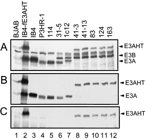
FIG. 2.
EBNA3 protein expression in LCLs infected with EBNA3AHT-expressing EBV recombinants. Immunoblots were incubated with EBV-immune human serum (A), sheep anti-EBNA3A polyclonal antibodies (B), or rabbit anti-estrogen receptor α polyclonal antibodies (C). Lane 1, lysates from EBV-negative BJAB human B lymphoblasts as a negative control; lanes 2 to 4, lysates from IB4 LCLs stably transfected with a FLAG-tagged EBNA3AHT (IB4-fE3AHT), IB4 cells, and P3HR-1 cells, respectively; lanes 5 and 6, lysates from LCLs 114 and 31-5, with type 2 wild-type EBNA3A-infected LCLs; lane 7, type 1 wild-type EBNA3A-infected LCL 1c12; lanes 8 to 12, five EBNA3AHT-infected LCLs. All LCLs, except for 114, expressed type 1 EBNA3B (E3B). All five EBNA3AHT-infected LCLs expressed a large EBNA3AHT (E3AHT) fusion protein and did not express wild-type 1 or 2 EBNA3A (E3A). Type 2 EBNA3C (E3C) is just below a background control band and was faintly detected by this EBV-immune human serum.

