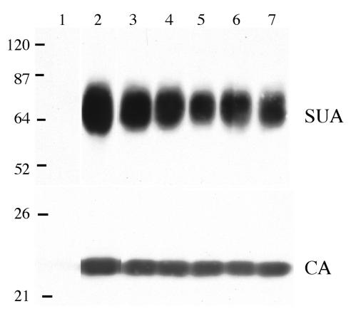
FIG. 4.
Western immunoblot analysis of the levels of SU glycoprotein in wild-type and mutant virions. Virions from day-16-infected DF-1 culture supernatants were pelleted, and the proteins were denatured, separated by SDS-12% PAGE, and transferred to a nitrocellulose membrane. A Western immunoblot containing the pelleted virus from 5 ml of supernatant was probed with an anti-subgroup A SU monoclonal antibody (SUA), and the bound protein complexes were visualized by chemiluminescence. A Western immunoblot containing the pelleted virus from 1 ml of supernatant was probed with anti-ALV CA sera (CA), and the bound protein complexes were visualized by chemiluminescence. In both immunoblots, proteins were analyzed from uninfected DF-1 cells (lane 1) and from DF-1 cells infected with either wild-type (lane 2), W141G K261E (lane 3), W145R K261E (lane 4), W141G (lane 5), K261E (lane 6), or W145R (lane 7) virus. Molecular sizes (in kilodaltons) are given on the left.

