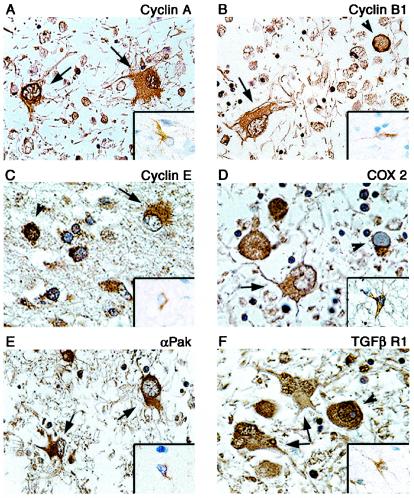
FIG. 3.
Immunohistochemical analysis of cellular proteins in demyelinated plaques of PML and normal brain tissue. Paraffin-embedded sections of brain tissue lesions from a patient with PML or from normal brain were analyzed for the expression of cyclin A (A), cyclin B1 (B), cyclin E (C), Cox-2 (D), PAK2 (E), and TGFβR1 (F). Insets show staining of normal brain tissue depicting a representative astrocyte with various antibodies as indicated in each panel. Bizarre astrocytes exhibit cytoplasmic immunoreactivity for cyclin A, cyclin B1, cyclin E, Cox-2, and PAK2 (arrows). Bizarre astrocytes exhibit cytoplasmic and punctate nuclear immunoreactivity when tested with an antibody for TGFβR1 (arrows). Oligodendrocyte inclusion bodies show cytoplasmic or perinuclear immunoreactivity for cyclin E, PAK2, TGFβR1, and Cox-2 and nuclear immunoreactivity for cyclin A, cyclin E, and TGFβR1 (arrowheads). Immunohistochemistry was performed by using the ABC Vector Elite system and was detected with DAB chromogen as described previously (14). All panels, original magnification ×1,000.

