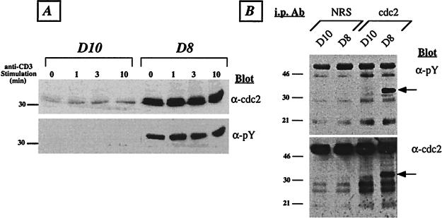
Figure 2.
Lck-deficient T cells express increased levels of predominantly tyrosine-phosphorylated cdc2 kinase. (A) T cells were activated by anti-CD3 cross-linking for the indicated times. Whole-cell lysates were prepared, and ≈50 μg of total protein was run per lane on 9% SDS/PAGE and immunoblotted with anti-cdc2 antibodies (Upper). Subsequently, the membrane was stripped and reprobed with 4G10 (anti-phosphotyrosine) mAb (Lower). The position of the 30-kDa molecular-mass standard is indicated. (B) Resting D10 and D8 cells were lysed in Nonidet P-40 containing lysis buffer, and ≈500 μg of total protein was immunoprecipitated with either normal rabbit serum (NRS) or cdc2-specific serum. The precipitates were resolved on 12.5% SDS/PAGE, transferred to poly(vinylidene difluoride) membrane, and immunoblotted wth 4G10 anti-phosphotyrosine(α-pY) mAb (Upper). Subsequently, the blot was stripped and reprobed with anti-cdc2 antiserum (α-cdc2; Lower). The molecular mass of standards is given in kDa. The cdc2 band is indicated by an arrow. The results are representative of five (A) and two (B) independent experiments.

