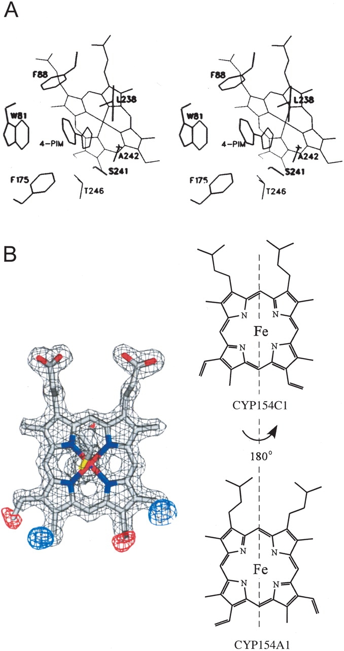
Figure 3.

Substrate binding site and heme orientation in CYP154A1. (A) Stereo view of 4-phenylimidazole binding in the active site of CYP154A1. (B) Heme orientation acquired from molecular replacement model CYP154C1. Fragments of Fo-Fc map (red and blue) indicate the requirement of a 180° heme rotation along the axis defined by the α- and γ-meso carbons, as schematically illustrated in the right part of the figure. Fragments of electron density from 2Fo-Fc composite omit map are contoured at 1.6σ (gray), from Fo-Fc map at −3.4σ (red), and 3.4σ (blue). The colors of the atoms are as follows: oxygen and iron, red; nitrogen, blue; sulfur, yellow; carbon, gray.
