
Figure 1.
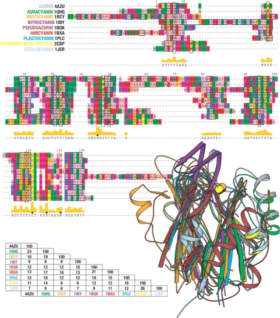
A structural alignment of nine members of the cupredoxin family. The alignment is based on a VAST (Gibrat et al. 1996) alignment, but has been manually improved. The coloring of the alignment is according to the zappo coloring scheme and was created using Pfaat (Johnson et al. 2003). The red boxes indicate β-strands and the blue boxes α-helices, as provided by dssp (Kabsch and Sander 1983). The black dashed vertical boxes indicate structurally conserved residues within the protein core. Stars indicate the position of the metal ligands. Sequence identities were calculated using Pfaat. The images were made using Molscript (Kraulis 1991).