
Figure 2.
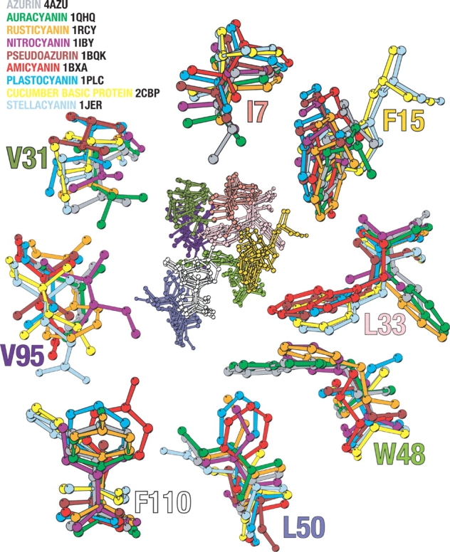
The superimposed hydrophobic core cluster of the nine different cupredoxins is seen in the center of the figure. Surrounding it are enlargements of the residues depicted in the central figure. The enlarged residues are colored according to the legend. The numbering used is according to that of P. aeruginosa azurin. Residues L33 and W48 have been enlarged together, not affecting their orientation with respect to one another. The side chain of these positions occupies the same spatial position with either position 33 or position 48 having a big bulky side chain, never both. In position 15 the case is similar; here the side chain filling the position comes from residues in another strand in stellacyanin, and basic cucumber protein compared to the other cupredoxins. The image was created using Molscript (Kraulis 1991).