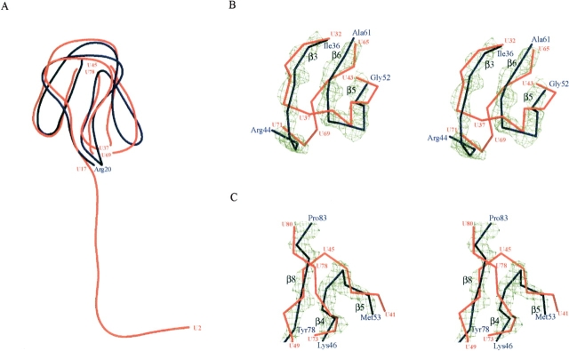
Figure 2.
Superposition of ribosomal protein L27 backbone representations from T. thermophilus (blue) and D. radiodurans (red). The blue residue numbers are for T. thermophilus and the red numbers are for D. radiodurans. (A) Overall structures of protein L27. Two pairs of Cα atoms (U37, U69, and U45, U78) were removed from the D. radiodurans protein L27. (B,C) Stereo showing the structural differences between the proteins L27 from T. thermophilus and D. radiodurans. The 2Fobs – Fcalc maps of the main-chain atoms of β8–β4–β5 (B) and β3–β6–β5 (C) are contoured at 2σ. The electron density surface was prepared by the program CONSCRIPT (Lawrence and Bourke 2000).

