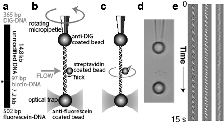
Figure 1.
Experimental design. (a) The molecular construct contains three distinct attachment sites and a site-specific nick (asterisk), which acts as a swivel. (b) Each molecule was stretched between two antibody-coated beads using a dual-beam optical trap. A rotor bead was then attached to the central biotinylated patch. The rotor was held fixed by applying a fluid flow, and the micropipette was twisted to build up torsional strain in the upper segment of the molecule. (c) Upon releasing the flow, the central bead rotated to relieve the torsional strain. (d) Video of the rotating bead (Supplementary Movies 2, 3) was analyzed to track cumulative changes in angle. Horizontal sections (red dashed line) of successive video frames can be stacked (e) to allow visualization of the helical path of the bead in space and time. (Left to right) Traces of 920 nm, 760 nm and 520 nm rotor beads.

