
Figure 1.
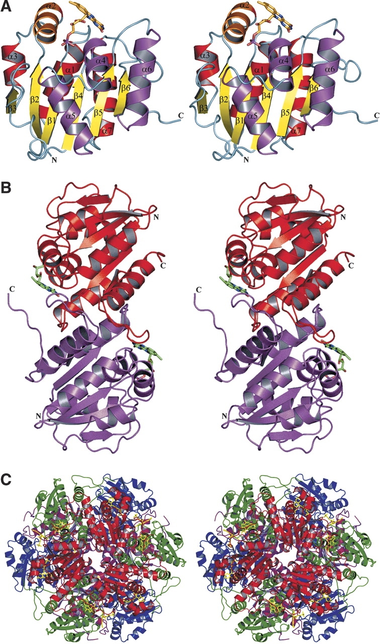
Structure of Pad1 decarboxylase. (A) Monomer: The central β-sheet is colored in yellow and the helices on one side of the β-sheet are shown in red, while on the other side they are in magenta. The helix on the top of the sheet and perpendicular to the strands is shown in orange. The FMN cofactor is shown as a stick model. (B) Pad1 dimer: The dimers associate tightly through interactions of the loops 121–127 and 146–158, helix α6 (128–140) and strand β6 (143–145) in one molecule with their counterparts in the second molecule. (C) Pad1 dodecamer: The four molecules within the asymmetric unit are colored blue, magenta, green, and red. This figure was prepared using the program PyMOL (http://www.pymol.org).