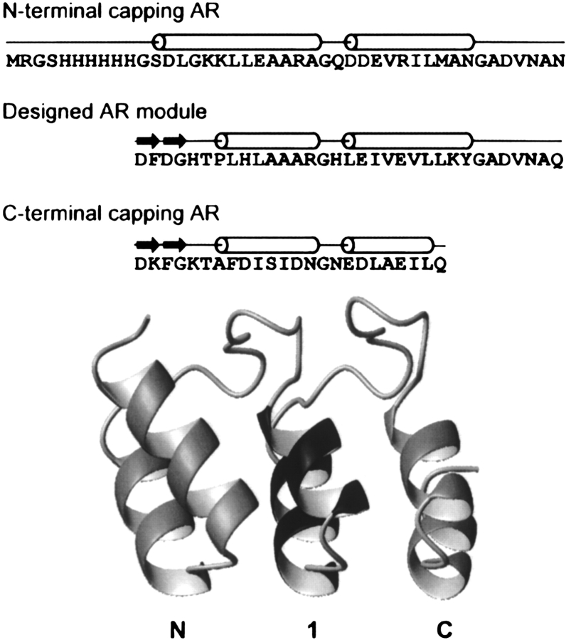
Figure 1.
The E_5 protein. After the N-terminal His tag, the sequence of the three ankyrin repeats are shown on separate lines. For orientation, the secondary structure elements are indicated above the sequence. Cylinders, α-helices; arrows, β-turns. In the structural model, the flanking capping repeats are shown in light gray, while the central consensus repeat is in dark gray. The N1C AR protein model was generated using homology modeling with Insight II (Accelrys) and the crystal structures of GABPb1 (PDB ID: 1AWC; Batchelor et al. 1998), E3_5 (1MJ0; Kohl et al. 2003), 3ANK (1N0Q; Mosavi et al. 2002), and 4ANK (1N0R; Mosavi et al. 2002) as templates. The picture was created using MOLMOL (Koradi et al. 1996).

