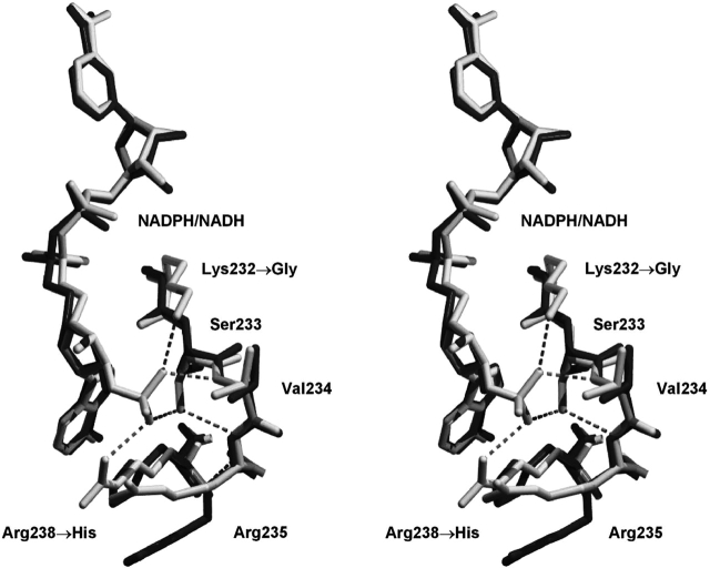
Figure 1.
Relaxed stereo diagram of the overlay of F22Y/K232G/R238H/A272G 2,5-DKGRA (dark gray) with wild type (1A80; light gray). The view is toward the adenine of the bound NADPH/NADH cofactor and provides structural details of the region involving the K232G and R238H mutations. The hydrogen bonding interactions of the 2′ adenosine phosphate group (of the NADPH bound in the wild-type structure) are indicated.

