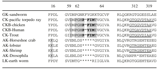
Table 1. Multi-sequence phosphagen kinase alignment in three regions.
Numbering is according to horseshoe crab arginine kinase. This alignment near 60–65 differs from prior ones, being based upon alignment of the horseshoe crab and pacific ray atomic structures (Zhou et al. 1998; Lahiri et al. 2002). Conserved lysine 16 (in arginine kinase) is shaded, as are sequence differences in the N-terminal domain specificity loop (between residues 59 and 63), with the creatine kinase insertion highlighted in bold. Asterisks represent imposed alignment gaps.

