
Figure 1.
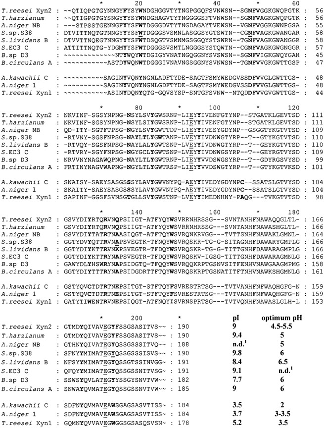
Sequence alignment of family 11 xylanases. The three acidophilic enzymes are from Aspergillus kawachii (XynC; Ito et al. 1992), Aspergillus niger (Xyl1; Krengel et al. 1996), and Trichoderma reesei (XYN1, Torronen et al. 1992). The so-called “alkaline” enzymes are from Trichoderma reesei (XYN2; Torronen et al. 1992), Trichoderma harzianum (Wong and Saddler 1992), Aspergillus niger (XynNB; Krengel and Dijkstra 1996), Streptomyces sp. S38 (Xyl1; Georis et al. 2000), Streptomyces lividans (XynB; Kluepfel et al. 1990), Streptomyces EC3 (xylanase C; Mazy-Servais et al. 1996), Bacillus D3 (Harris et al. 1997), and Bacillus circulans (XynA; Yang et al. 1989). The residues referred to in the text are in bold, the catalytic Glu residues underlined, and the mutated residues in bold and underlined. The individual numbering of each enzyme is shown. In the text, the standard numbering resulting from the alignment is used consistently. n.d.: Not determined.