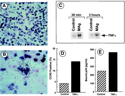
Figure 6.
MAg stimulates TNFα expression. LN from HIV-1-infected (A) and HIV-1 plus MAC-infected (B) subjects were processed for ISH with a TNFα antisense probe. Sense probes were negative. (C) Monocytes were stimulated in vitro with MAg (25 μg/ml) and the isolated RNA probed for TNFα by Northern hybridization. (D) Monocytes were exposed to TNFα (10 ng/ml) overnight before FACS analysis for CCR5. (E) Monocytes were treated with TNFα for 4 hrs and exposed to HIV-1 at TCID50 = 4 × 10³ for 30 min. Cells were washed, lysed, and assayed for p24 levels by ELISA.

