
Figure 7.
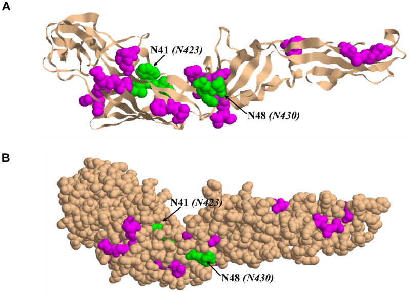
HCV E2 homology model based on the Tick Borne Encephalitis Virus envelope glycoprotein E (TBEV) structure (PDB id: 1SVB): (A) Cartoon representation of E2 glycoprotein showing the location of the glycans attached to the protein. The nine sites of glycosylation found to have attached only high mannose type sugars are highlighted in magenta, and the two sites found to have complex type glycans attached are shown in green. (B) Another view of the E2 glycoprotein illustrated in space filling representation. The same colors as described above were used to indicate the location of the glycans on E2.