Figure 1.
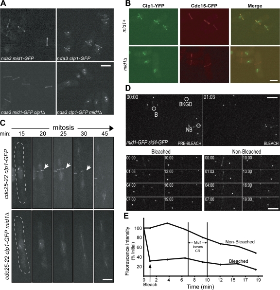
Clp1 depends on Mid1 for localization to the CR. (A) Live-cell images of nda3-KM311 mid1-GFP, nda3-KM311 mid1-GFP clp1Δ, nda3-KM311 clp1-GFP, and nda3-KM311 clp1-GFP mid1Δ cells after incubation at 18°C for 7 h. (B) Live-cell images of nda3-KM311 clp1-YFP cdc15-CFP and nda3-KM311 clp1-YFP cdc15-CFP mid1Δ cells after arrest by incubation at 18°C for 7 h. Over 100 cells were examined for each strain. (C) Time-lapse confocal microscopy of live cdc25-22 clp1-GFP and cdc25-22 clp1-GFP mid1Δ cells during mitosis. Exponentially growing cells were incubated at the restrictive temperature (36°C) for 3.5 h, and then released to the permissive temperature (25°C) for 15 min. Indicated time points are from time of release. Arrows indicate Clp1 ring. Images are from Videos 1 and 2 (available at http://www.jcb.org/cgi/content/full/jcb.200709060/DC1). (D and E) Representative images (D) and fluorescence recovery curves (E) for Mid1-GFP FRAP in mid1-GFP sid4-GFP cells. Sid4-GFP signal determined mitotic stage and vertical lines indicate the window when Mid1 begins to dissociate from the CR. B, bleach region; NB, nonbleached region (used to correct for overall bleaching); BKGD, background region (used to correct for overall bleaching). Images are from Video 3 (available at http://www.jcb.org/cgi/content/full/jcb.200709060/DC1). Bars, 5 μm.

