Figure 3.
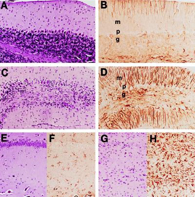
Doxycycline prevented neuronal loss, vacuolation, and gliosis in Tg(tTA:PrP)3 mice inoculated with RML prions. (A, B, E, and F) Tg(tTA:PrP)3 mice treated with oral doxycycline (2 mg/ml) and sacrificed at 200 days postintracerebral inoculation with RML prions. (C, D, G, and H) Tg(tTA:PrP)3 mice untreated with doxycycline and sacrificed at 70 days postintracerebral inoculation with RML prions when they showed multiple signs of CNS dysfunction; the first sign of neurologic disease was ataxia at 50 days after inoculation. (A, C, E, and G) Hematoxylin and eosin-stained brain sections. (B, D, F, and H) α-GFAP-immunostained brain sections. (A–D) Views of cerebellar molecular (m), Purkinje cell (p), and granular (g) layers. (E–H) Views of the hippocampus CA1 pyramidal cell layer (aligned in the upper part of Insets) with underlying stratum radiatum. Focal loss of Purkinje cells and granule cells was observed in the cerebellum of the ill Tg(tTA:PrP)3 mice and was accompanied by low grade vacuolation. Reactive astrocytic gliosis was demonstrated by the α-GFAP immunostaining that revealed moderate to severe gliosis in the granular and molecular layers (D) (Bergmann’s gliosis). In the hippocampal CA1 region most neuronal cell bodies were lost, and low grade vacuolation was apparent together with severe astrocytic gliosis (H).

