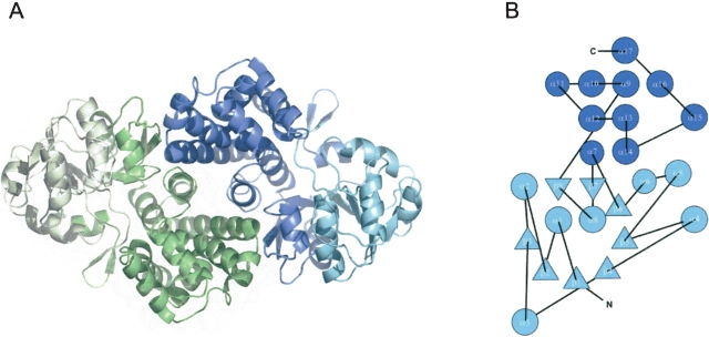
Figure 4.
Structure of NADP+-dependent G3PDH from A. fulgidus. (A) Ribbon diagram of the G3PDH dimer when viewed perpendicular to the twofold noncrystallographic axis. One monomer is colored in green, one in blue. The N-terminal dinucleotide binding domains are shown in light green and in light blue; the C-terminal helix domains are shown in dark green and dark blue. The figure was produced with MOLSCRIPT (Kraulis 1991) and Raster3D (Merritt and Murphy 1994). (B) Fold topology diagram of the G3PDH monomer.

