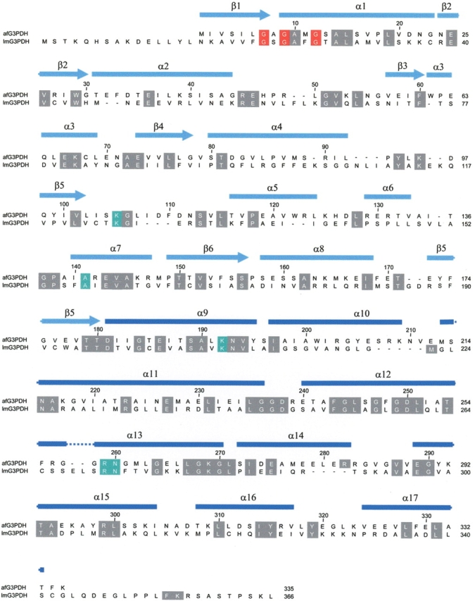
Figure 5.

Alignment of the primary structures of G3PDH from A. fulgidus and L. mexicana. The residues, which are involved in substrate binding including the dinucleotide binding site GXGXXG (residues 7,9,12), are depicted in red. The conserved substrate binding sites are highlighted in green. The secondary structure assignment of G3PDH from A. fulgidus is shown above the sequence alignment.
