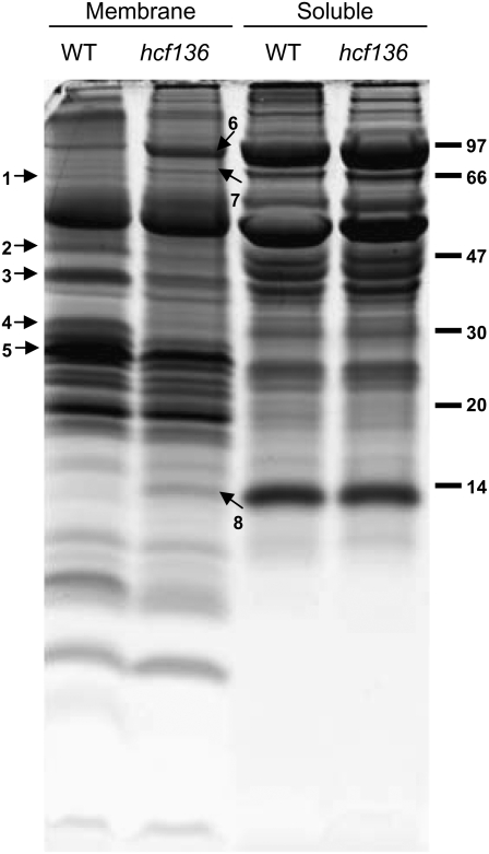
Figure 5.
Electrophoretic pattern of wild-type (WT in the image) and hcf136 proteins obtained by SDS-PAGE (tricine 12%) and stained with Sypro Ruby fluorescent dye. Total thylakoid membrane vesicles were isolated on Percoll cushions and then treated with a Dounce homogenizer followed by differential ultracentrifugation to collect membrane and soluble fractions. Bands displaying strong differential accumulation were excised and proteins digested and analyzed by MALDI-TOF MS PMF. Identified proteins are: (1) FtsH1 (TC292243); (2) CP47 (NP_043049.1); (3) OEC33-like (TC279249); (4) PSII-D2 (NP_043009.1); (5) LHCII-1 (TC286614); (6) PPDK (TC286559); (7) cpHsp70 (TC293193), and (8) RBCS (TC286731). These proteins are labeled with numbered arrows. Protein markers in kilodaltons are indicated on the right.

