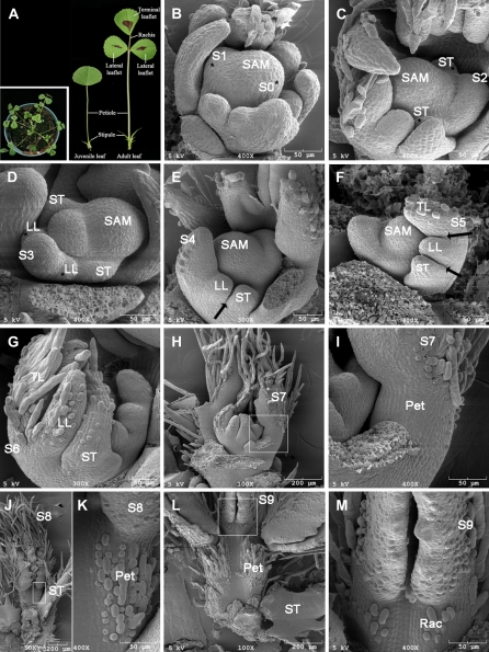
Figure 1.
The ontogeny of compound leaf development in wild-type M. truncatula. A, Morphology of M. truncatula ‘Jemalong’ A17. Shown in inset were juvenile (left) and adult (right) leaves. B to M, SEM analysis of compound leaf development. B, Sites of incipient leaf primordia were specified at the periphery of the SAM at S0, albeit no morphological changes were visible at this stage. At S1, a common leaf primordium was initiated as a strip of cells outgrown along the periphery of SAM (asterisk). C, A pair of stipule primordia (ST) was initiated from the proximal end of the common leaf primordium at S2. D, At S3, a pair of lateral leaflet primordia (LL) emerged between the stipule and common leaf primordia. E, Boundaries (arrow) between the stipule and lateral leaflet primordia formed, and the common leaf primordium differentiated into a terminal leaflet primordium (TL) as indicated by development of trichomes from the abaxial surface at S4. F, At S5, boundaries (arrows) formed between the lateral and terminal leaflet primordia. G, While trichomes developed from the abaxial surface of both stipule and lateral leaflet primordia, the terminal leaflet primordium folded as a result of outgrowth of the abaxial surface at S6. H, At S7, a petiole primordium (Pet) formed between the stipule and lateral leaflet primordia. I, A close-up view of H. J, The lateral and terminal leaflet primordia folded due to outgrowth of the abaxial surface at S8. Trichomes developed from the adaxial surface of the petiole primordium at this stage. K, A close-up view of J. L, A rachis primordium (Rac) formed between the lateral and terminal leaflet primordia and trichomes developed from its adaxial surface at S9. M, A close-up view of L. Scale bars, 50 or 200 μm as indicated.

