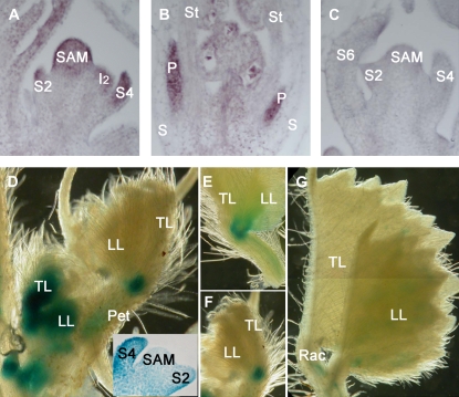
Figure 5.
Expression pattern of SGL1 gene. A to C, RNA in situ hybridization analysis of SGL1 gene expression. A, SGL1 gene expression was detected in SAM, developing leaf primordia (S2 and S4), and I2. B, SGL1 gene expression was detected in developing floral organs in S7 flowers of wild-type plants. P, Petal; S, sepal; St; stamen. C, SGL1 sense probes were used as a negative control, no hybridization signal was detected in SAM and leaf primordia (S2, S4, and S6). D to G, SGL1:GUS histochemical staining pattern. D, GUS staining was restricted to the SAM (inset), developing leaf primordia at early stages (inset), vascular tissues of petioles, and the basal regions of leaflets at late stages. E and F, Close-up views of GUS staining patterns at the basal region of leaflets. G, GUS staining in mature leaves.

