Figure 7.
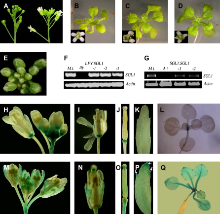
Genetic complementation of lfy mutants by SGL1 and comparison of expression patterns of LFY:GUS and SGL1:GUS in Arabidopsis. A, Inflorescence of homozygous lfy (left) and lfy transformed with LFY:SGL1 (right). B to D, Two-week-old wild-type (B), lfy LFY:SGL1 (C), and lfy SGL1:SGL1 (D) seedlings; insets, mature flowers. E, Morphology of lfy mutant flowers. F, RT-PCR analysis of SGL1 expression in wild-type M. truncatula (lane 1), lfy (lane 2), and three independent transgenic lfy lines transformed with LFY:SGL1 (lanes 3–5). G, RT-PCR analysis of SGL1 expression in M. truncatula (lane 1), Arabidopsis (lane 2), and two independent transgenic lfy lines transformed with the SGL1 genomic sequence (SGL1:SGL1; lanes 3 and 4). An Actin gene was used as an internal loading control (F and G; bottom). H and L, Histochemical staining of LFY:GUS in mature and young seedlings. Strong GUS staining was restricted to the base of flowers, vascular tissues of inflorescence stems, and the shoot apex but not of pedicels and leaves. M to Q, Histochemical staining of SGL1:GUS in mature and young seedlings. Strong GUS staining was detected in style, but not at the base of flowers, and in vascular tissues of inflorescence stems, pedicels, sepals, carpel, cauline, and rosette leaves, and the shoot apex. In cauline and rosette leaves, a high level of GUS staining was localized to the distal margins (arrows).

