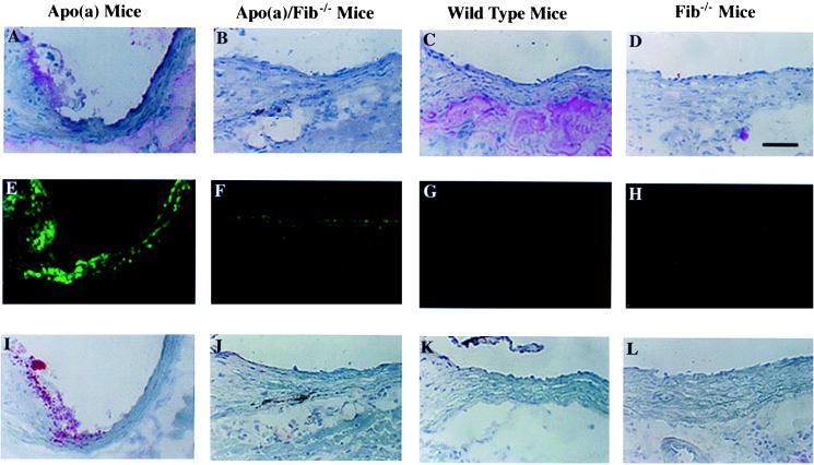
Figure 1.
Immunofluorescent and histochemical staining of fibrin(ogen) and apo(a) accumulation and lipid deposition in proximal sections of mouse aorta after 2 months of an atherogenic diet. Representative adjacent sections are shown for the three different types of staining. (A–D) Immunohistochemical staining of fibrin(ogen) in apo(a), apo(a)/Fib−/−, wild-type, and Fib−/− mice, respectively. (E–H) Immunofluorescent staining of apo(a) accumulation. (I–L) Oil-red O staining for fatty streak lesions. Note more lipid is in the apo(a) mice, including the edge of the valve and the vessel walls. Fibrin(ogen) staining is essentially colocalized with apo(a) accumulation and fatty streak lesions. (Scale bar equals 50 μm.)

