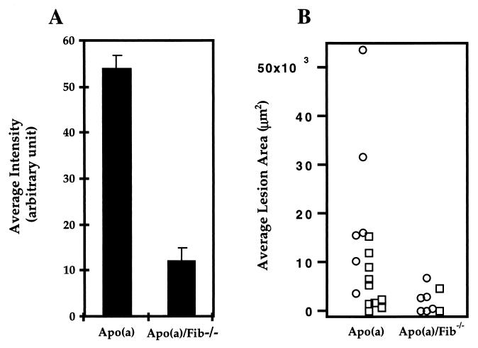
Figure 2.
Quantitative analyses of immunofluorescent-stained apo(a) accumulation and oil-red O-stained lipid lesions in the proximal aorta of apo(a) and apo(a)/Fib−/− mice all maintained on high-fat diet. (A) Group mean of average vessel wall intensity [arbitrary units (1–250)/area (number of pixels)] in apo(a) (n = 11) and apo(a)/Fib−/− mice (n = 8). The mean of average intensity for apo(a) mice is 53.7 ± 3.3; for apo(a)/Fib−/− mice it is 11.8 ± 2.2 (P = 1.17 × 10 −8). The staining was performed as one batch. (B) Average lipid lesion area per section per mouse (females, ○; males, □). The mean of the average lesion area per section per mouse is 11,584 ± 3,465 μm2 for the apo(a) mice and 2,161 ± 889 μm2 for the apo(a)/Fib−/− mice. The median is 7,865 μm2 (range: 0–53,560) for the apo(a) mice and 1,560 μm2 (range: 0–6,760) for the apo(a)/Fib−/− mice (P = 0.02).

