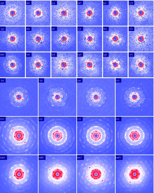
Figure 4.
End-on microdiffraction patterns of quick-frozen DLM myofibrils from pupae at various developmental stages and from adult flies. (a–z) Diffraction from myofibrils from pupae. (a–c) 23–27 h APF showing speckle-like scattering which often forms a peak at a radius smaller than at 1/50 nm−1, typically 1/57 nm−1. In (c), the periodical intensification of scattering is software detected at 60° intervals in the scattering angle expected for the 1,0 reflection (1/40–1/45 nm−1; boxes; see text for details). (d–f) 42–45 h APF. (g–l) 58–61 h APF. (m,n) 76–77 h APF. Shown are among the patterns which show software-detected intensification at 60° intervals in the scattering angle expected for the 1,0 reflection (boxes). In (m,n), the hexagonal arrangement of the 1,0 peaks are visibly clear, but the 2,0 reflections are still obscure. (s–v) 87 h APF. Both 1,0 and 2,0 reflections are clearly observed, as well as some higher-order reflections. (w–z) More than 100 h APF (about to eclose, age estimated from appearance). (aa–ad) Adults (age not recorded). In these patterns, the features of hexagonal myofilament lattice are clearly observed (see Iwamoto et al. 2006a). Isotropic scattering has been subtracted from patterns (a–v).

