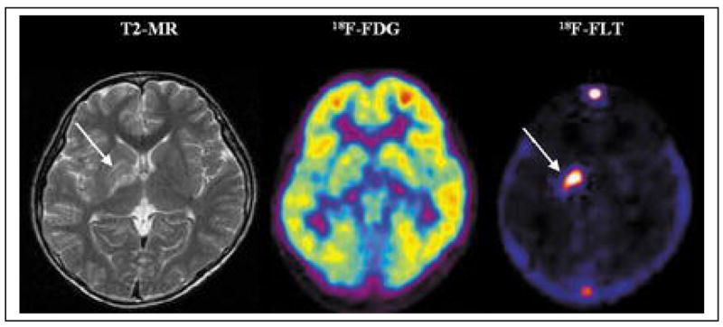
Figure 1. Comparison of FDG and FLT.

An 11-year-old male with a germ cell tumor of the basal ganglia. MRI shows subtle changes (arrow) in the right basal ganglia. On FDG-PET, the right basal ganglia lesion shows slightly decreased uptake compared with the contralateral basal ganglia but increased uptake compared with normal white matter. 3′-Deoxy-3′-[18F]fluorothymidine (FLT)-PET, however, reveals intensely increased uptake, suggesting the presence of a malignant tumor (arrow). Based on the FLT-PET results, a stereotactic biopsy was performed in the right putamen. could be performed in the right basal ganglia (63).
From: Choi SJ, Kim JS, Kim JH, et al. [18F]3′-deoxy-3′-fluorothymidine PET for the diagnosis and grading of brain tumors. Eur J Nucl Med Mol Imaging. Jun 2005;32(6):653–659.
