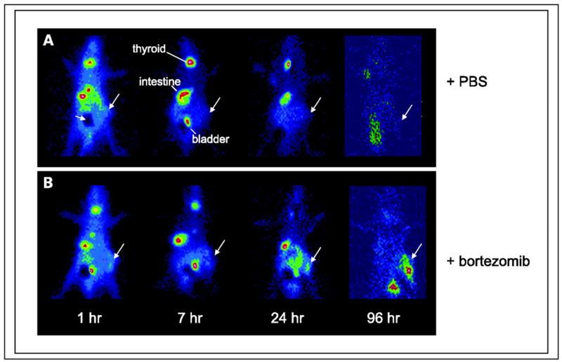
Figure 3. Viral TK induction.

Time course of uptake of [125I]FIAU by Burkitt’s lymphoma xenografts [EBV(+) Akata] following treatment with bortezomib as assessed by planar gamma scintigraphy in vivo. Large arrows, tumors. The dark area (A, small arrow) represents lead shielding of bladder to improve the dynamic range of the images. Each animal has one tumor placed in the hind limb. A, no tumor uptake is evident in animals pretreated with PBS only (control). B, tumors are visualized at later time points in the pretreated animals (2 μg/g bortezomib).
From: Fu DX, Tanhehco YC, Chen J, et al. Virus-associated tumor imaging by induction of viral gene expression. Clin Cancer Res. Mar 1 2007;13(5):1453–1458.
