Figure 4.
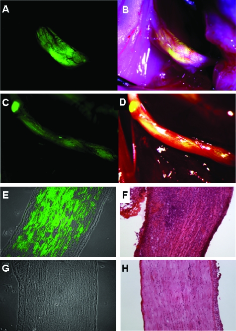
NV1066 selectively localizes areas of nerve infiltration by prostate and pancreatic carcinoma cells. Fluorescence image (A) and overlay image of prostate carcinoma (DU145)-derived tumor (B) and pancreatic carcinoma (MiaPaCa2)-derived tumor (C and D). Fluorescence microscopy (E) and corresponding hematoxylin and eosin image (F) of a sciatic nerve invaded by prostate carcinoma cells (PC3) and treated with NV1066. Fluorescence microscopy (G) and corresponding hematoxylin and eosin image (H) of a sciatic nerve without tumor treated with NV1066. Enhanced green fluorescent protein (eGFP) expression was found only in nerves infiltrated by cancer but not in normal nerves. Panels A to D are stereomicroscopic images. Panels E to H are microscopic images original magnifications, x10).

