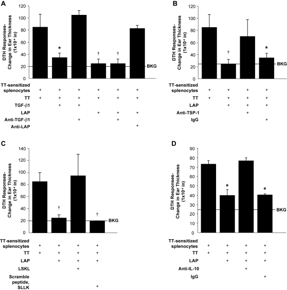
Figure 3. LAP is able to suppress in vivo cellular inflammation similar to TGF-β1 through interactions with thrombospondin-1.
A transfer DTHR assay was performed with antigen (tetanus toxin) and tested agents (TGF-β1, rhLAP and anti-TGF-β1, LAP or TSP-1). C57BL/6 mice were sensitized to tetanus toxin (TT) and then tested to confirm they had developed a TT-induced DTHR. DTHR was induced in the pinnae of naïve C57BL/6 mice with simultaneous injection (35 µl) of antigen and splenocytes (8×10⁶ cells/condition) harvested from antigen-sensitized mice. DTHR was quantified by measurement of ear thickness 24 hours after injection. (A) LAP (10 pg) and TGF-β1 (5 ng) were tested for the ability to reduce DTHR when used alone, with anti-TGF-β1 antibodies (1 µg) or anti-LAP antibodies (1 µg) (*p<0.02 versus tetanus sensitized splenocytes + TT; †p<0.01 versus tetanus sensitized splenocytes + TT, n = 5). Similar results were observed when this assay was repeated using C57BL/6 mice that had rejected a cardiac allograft (DBA/2->C57BL/6) as a model of transfer DTHR. (B) Anti-TSP-1 antibody or isogenic control IgG (all at 1 µg) were tested for their ability to interfere with rhLAP suppression of TT-induced DTHR (*p<0.02 versus tetanus sensitized splenocytes + TT; †p<0.01 versus tetanus sensitized splenocytes + TT, n = 3). (C) Transfer DTHR assay was performed with antigen, rhLAP and the inhibitory peptide, LSKL or its scrambled control SLLK (20 µM) (†p<0.01 versus tetanus-sensitized splenocytes + TT, n = 2). (D) To determine if LAP used an IL-10 dependent pathway to inhibit the DTHR (as previously published) the DTHR transfer assay was repeated with anti-IL-10 antibodies and isotype control. (1 µg each) (*p<0.01 vs. tetanus-sensitized splenocytes + TT, n = 3) All bars represent the mean±SEM. DTHR, delayed-type hypersensitivity response; Ag, Antigen; BKG, background ear girth.

