
FIGURE 1.
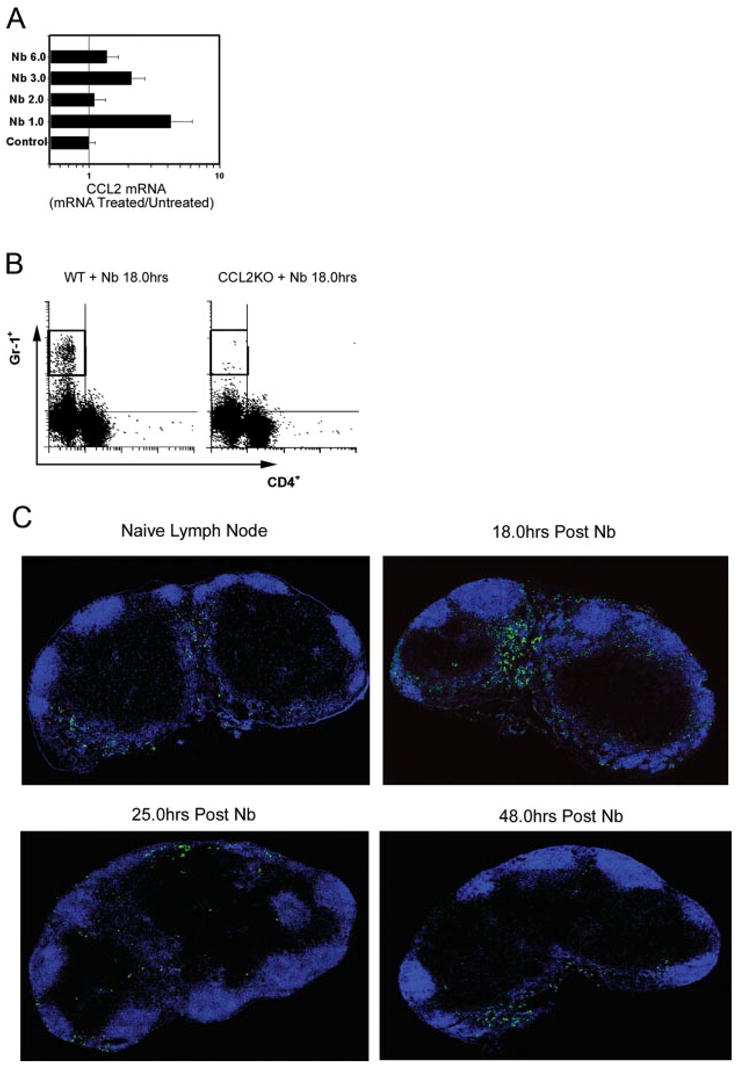
CCL2-dependent Gr-1bright cell infiltration of the draining lymph node shortly after N. brasiliensis inoculation. A, BALB/c mice (5/treatment group) were intracutaneously inoculated with 500 N. brasiliensis L3. On days 1, 2, 3, or 6 after inoculation, draining CLN were removed, RNA purified, and analyzed for CCL2 mRNA using real-time quantitative fluorogenic RT-PCR. Fold changes are expressed relative to the untreated BALB/c control. B, CCL2KO and WT control mice (5/treatment group) were inoculated with N. brasiliensis L3 at the ear. Draining CLN were collected 18 h later. CLN cell suspensions were prepared and stained for anti-Gr-1-PE and anti-CD4-FITC. C, BALB/c WT mice were inoculated intracutaneously in one ear with N. brasiliensis L3. Draining CLN were removed at 18, 24, or 48 h after inoculation, and also from untreated control mice. CLN frozen sections were fluorescently stained with anti-B220-Alexa Fluor 647 (B cells; blue) and anti-Gr-1-FITC (green). Individual × 400 digital images were tiled together to form a single high resolution composite image of the draining lymph node. These experiments were repeated twice with similar results.