
Figure 4. Hint/intein containing genes in N. vectensis are likely involved in neural patterning.
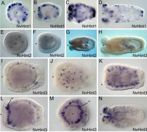
A-D. Expression of NvHint1 is punctate in the ectoderm and initiates in the early planula stage (A). Expression increases at mid-planula (B) and persists into the polyp (C,D). Expression is exclusively ectodermal, in scattered cells that are likely to be neural in origin due to their basal position. Expression is relatively uniform across the oral-aboral axis of the planula, but becomes more concentrated at both the oral and aboral ends of the polyp. E-H. Expression of NvHint2 is confined in a cell-type specific manner to body-wall endoderm and cells within the directive mesenteries. Expression is detected in a few scattered endodermal cells in the planula (E-F). In the polyp expression can be detected in both body-wall endodermal cells and within the directive mesenteries (G-H). I-N. Expression of NvHint3 is also ectodermal and begins in a punctate fashion in the planula (I-K). Expression in the early polyp becomes elevated in tentacular ectoderm as well as a ring of cells surrounding the mouth, which are likely to be components of the circumoral nerve ring (nr) (arrows, L, M). Expression persists mainly in the tentacular ectoderm with some scattered expression in the body wall ectoderm of the polyp (N). All embryo views are lateral, with the asterisk denoting the blastopore and future mouth, except A, E, M, which are oral views.